15 December 2021 : Case report
Rex Shunt for Portal Vein Thrombosis After Pediatric Living Donor Liver Transplantation
Yuji Soejima1ABDE*, Tomoaki Taguchi2DE, Toshiharu Matsuura2D, Makoto Hayashida2D, Toru Ikegami1BD, Tomoharu Yoshizumi1BD, Yoshihiko Maehara1DDOI: 10.12659/AOT.909493
Ann Transplant 2021; 26:e909493


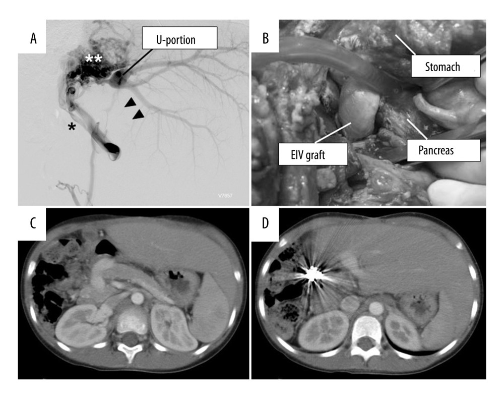
Figure 3 (A) Angiographic findings of portal vein thrombosis of Case 3. Note that the portal vein trunk is completely thrombosed (arrowheads) and a large collateral vein (*) with marked cavernous transformation (**) has formed. (B) Intraoperative view of the Rex shunt created between the SMV and the UP using the donor’s external iliac vein. Note the shunt graft is tunneled through the prepancreatic retropyloric route. (C, D) A computed tomography scan taken at 3.3 years after the procedure shows a patent Rex shunt graft (C) and the UP of the graft (D). EIV – external iliac vein; UP – umbilical portion of the portal vein.