19 June 2020 : Original article
Safety and Efficacy of Right Retroperitoneal Laparoscopic Live Donor Nephrectomy: A Retrospective Single-Center Study
Yaowen Fu1ABCEF, Yu Hu2ABCEF, Weigang Wang1ABCF, Baoshan Gao1ABCE, Gang Wang1ABCE, Xin Lian1ABC, Honglan Zhou1ABEG*, Yuantao Wang1AEGDOI: 10.12659/AOT.919284
Ann Transplant 2020; 25:e919284


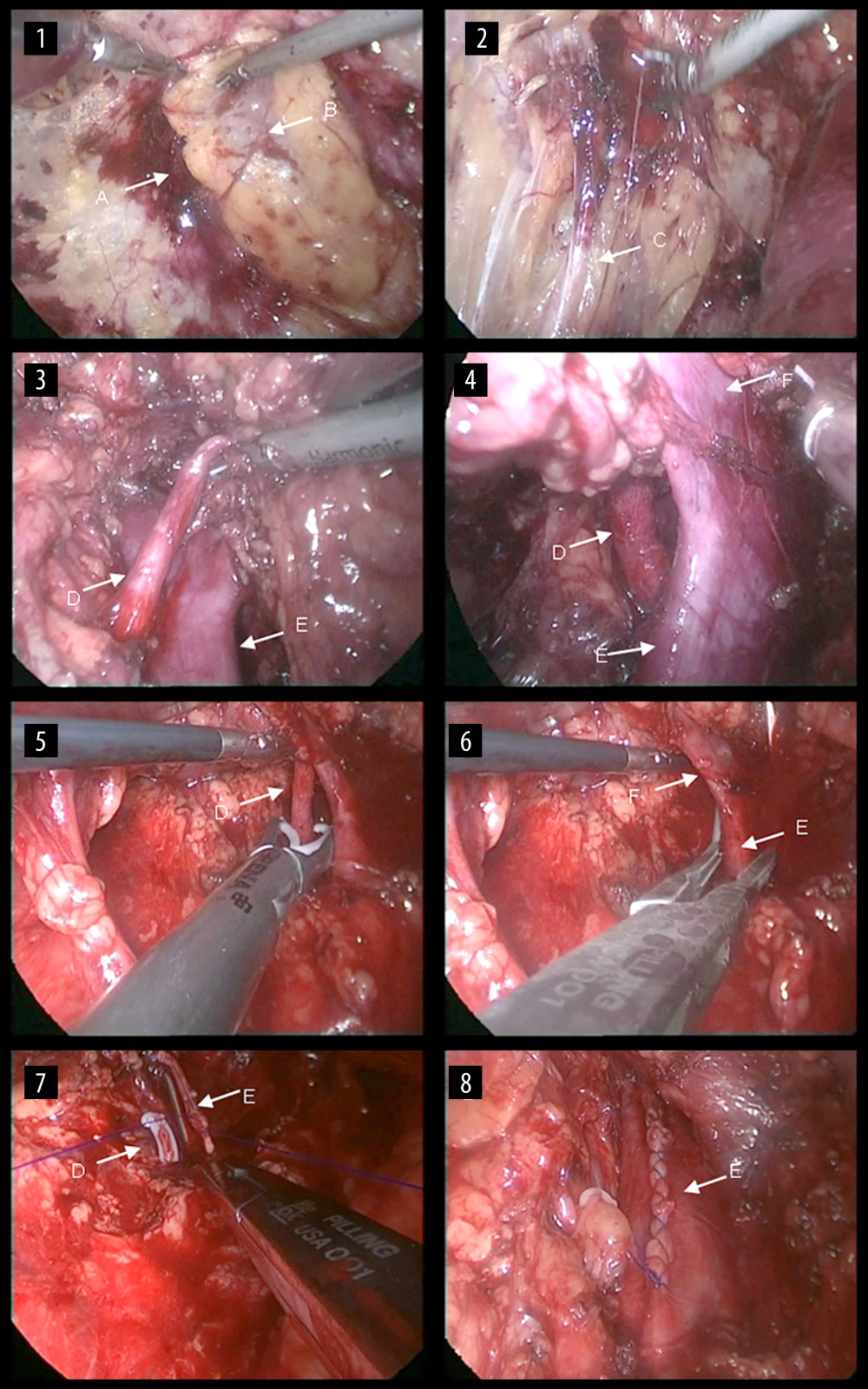
Figure 3 Surgical steps of right retroperitoneal laparoscopic live donor. nephrectomy. The renal fascia is opened (1), the ureter is fully dissociated (2), and then the right renal artery is dissected (3). The renal vein and the inferior vena cava are carefully dissected at their confluence (4). Then, the renal artery is clipped proximally (5), the renal vein is blocked at the confluence of the renal vein at the inferior vena cava and the renal vein is cut along with partial vena cava wall (6). The incision on the lateral wall of the inferior vena cava is then continuously sutured (7) and the vena cava after suturing is shown (8). A: the renal fascia; B: the right kidney; C: the ureter; D: the right renal artery; E: the inferior vena cava; F: the right renal vein.