22 September 2020 : Original article
Intensive Doppler Ultrasonography for Early Detection of Hepatic Artery Thrombosis After Adult Living Donor Liver Transplantation
Pi-Ling Chiang1ABCDEF, Yu-Fan Cheng1ADG, Tung-Liang Huang1BG, Hsin-You Ou1B, Chun-Yen Yu1B, Hsien-Wen Hsu2B, Wei-Xiong Lim2B, Chao-Long Chen3B, Chee-Chien Yong3B, Leo Leung-Chit Tsang1ABDEF*DOI: 10.12659/AOT.924336
Ann Transplant 2020; 25:e924336


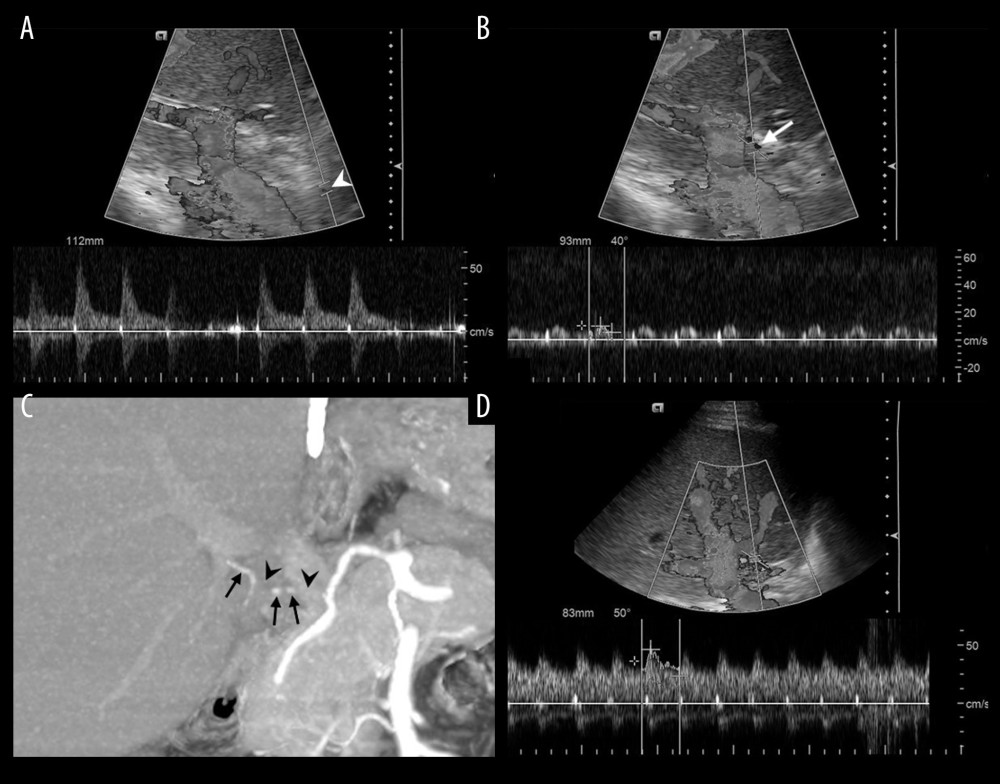
Figure 1 The 66-year-old male hepatocellular carcinoma patient received LDLT with right lobe graft. Right hepatic artery of the graft were anastomosed with the recipient’s right hepatic artery. An abnormal Doppler waveform was detected on post-LDLT day 12. (A) Doppler US demonstrated normal arterial curve at the pre-anastomotic artery (white arrowhead), and (B) tardus parvus waveform at post-anastomotic intrahepatic artery (RI=0.46, PSV=10.1 cm/s, white arrow). (C) Subsequent CTA on the same day demonstrated multifocal disruption of post-anastomotic artery (black arrowheads) and faint opacification of intrahepatic arterial filling (black arrows), suggesting partial occlusion. After IVT, (D) DUS on the next day demonstrated recovered arterial curve (RI=0.64, PSV=47 cm/s).