22 September 2020 : Original article
Intensive Doppler Ultrasonography for Early Detection of Hepatic Artery Thrombosis After Adult Living Donor Liver Transplantation
Pi-Ling Chiang1ABCDEF, Yu-Fan Cheng1ADG, Tung-Liang Huang1BG, Hsin-You Ou1B, Chun-Yen Yu1B, Hsien-Wen Hsu2B, Wei-Xiong Lim2B, Chao-Long Chen3B, Chee-Chien Yong3B, Leo Leung-Chit Tsang1ABDEF*DOI: 10.12659/AOT.924336
Ann Transplant 2020; 25:e924336


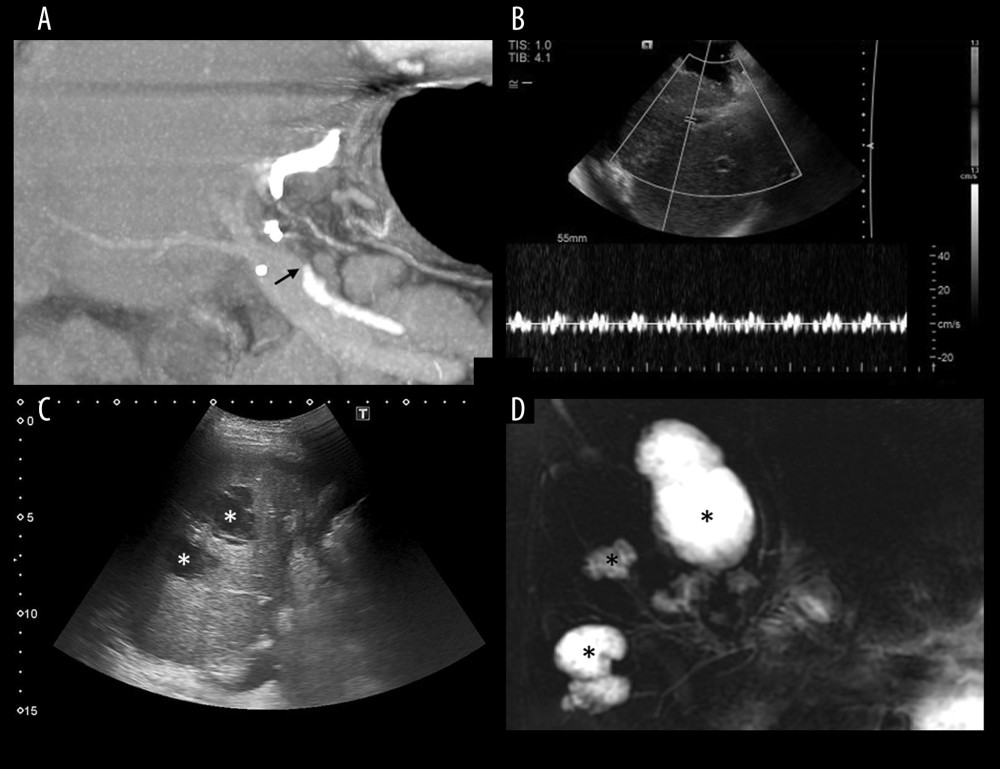
Figure 2 The 43-year-old male liver cirrhosis patient received LDLT with right lobe graft. Right anterior and right posterior hepatic arteries (RAHA and RPHA) of the graft were separately anastomosed with the recipient’s gastroepiploic artery and left hepatic artery. On post-LDLT day 1, DUS demonstrated complete absence of arterial wave of RAHA and RPHA (not shown). (A) CTA on the same day demonstrated disruption of periportal HA (black arrows) and non-opacification of intrahepatic arterial branches. (B) Intra-operation DUS on the same day demonstrated damped and irregular systolic upstroke and absent diastolic flow around the anastomotic site. Re-anastomosis of RAHA with gastroepiploic artery and IVT were done. Post re-anastomosis DUS showed recovered RAHA arterial blood flow (not shown in this picture). (C, D) Due to repeated arterial occlusion, multi-segmental non-anastomotic intrahepatic biliary strictures with bilomas (*) were noted 4 month later in US and MRCP. The patient received deceased donor liver transplantation 9 months later. The histologic findings of the LDLT allograft were ischemic necrosis, acute and chronic cholangitis, and multifocal abscess formation.