04 August 2020 : Original article
Inferior Vena Cava Constriction After Liver Transplantation Is a Severe Complication Requiring Individually Adapted Treatment: Report of a Single-Center Experience
Jan-Paul Gundlach1CDEF*, Rainer Günther2B, Marcus Both3BD, Jens Trentmann3BD, Jost Philipp Schäfer3BCD, Jochen T. Cremer4B, Christoph Röcken5BDE, Thomas Becker1DEG, Felix Braun1ABD, Alexander Bernsmeier1CDEDOI: 10.12659/AOT.925194
Ann Transplant 2020; 25:e925194


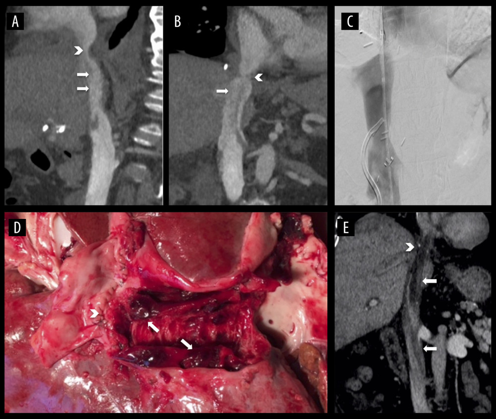
Figure 1 Radiologic and macroscopic finding of IVC thrombosis. (A–C): patient #1: CT scan (A, B) and cavography (C) of thrombus due to fibrotic stenosis (arrow heads) of suprahepatic IVC anastomosis; (D) patient #2 at autopsy with cranio-caudal opened IVC (from left to right); arrow head indicates suprahepatic IVC anastomosis; (E) CT scan showing a massive IVC thrombosis in patient #3 with arrowhead indicating stenotic suprahepatic IVC anastomosis. White arrows indicating thrombosis. Pictures A–C, and E are displayed in coronal view.