05 July 2022 : Original article
Cardiac and Pulmonary Histopathology in Baboons Following Genetically-Engineered Pig Orthotopic Heart Transplantation
Silvio H. Litovsky1BDE, Jeremy B. Foote2BCDE, Abhijit Jagdale3BE, Gregory Walcott4BE, Hayato Iwase3BDE, Mohamed H. Bikhet3BDE, Takayuki Yamamoto3BE, Christophe Hansen-Estruch3BE, Mohamed B. Ezzelarab5BE, David Ayares6AE, Waldemar F. Carlo7BE, Leslie A. Rhodes7BE, Jack H. Crawford8BE, Santiago Borasino8BE, Robert J. Dabal8BE, Luz A. Padilla8BE, Hidetaka Hara3BE, David K.C. Cooper3ABDEFG*, David C. Cleveland8ABDEFGDOI: 10.12659/AOT.935338
Ann Transplant 2022; 27:e935338


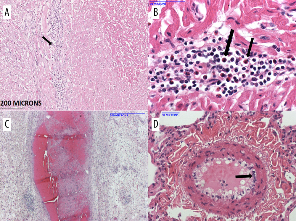
Figure 4 Histopathology of the pig heart (A–D) in B15416 at necropsy(A) The heart showed mild inflammation and fibrosis within the myocardial interstitium, and around the Purkinje fibers. Higher magnification showed a fair number of eosinophils in the infiltrate (B). The coronary arteries (C) exhibited significant mature fibrin thrombi, resulting in narrow intravascular lumens. Within the lungs (and heart, not shown), there were multiple medium-caliber arteries with endothelial activation (D, arrow).