05 July 2022 : Original article
Cardiac and Pulmonary Histopathology in Baboons Following Genetically-Engineered Pig Orthotopic Heart Transplantation
Silvio H. Litovsky1BDE, Jeremy B. Foote2BCDE, Abhijit Jagdale3BE, Gregory Walcott4BE, Hayato Iwase3BDE, Mohamed H. Bikhet3BDE, Takayuki Yamamoto3BE, Christophe Hansen-Estruch3BE, Mohamed B. Ezzelarab5BE, David Ayares6AE, Waldemar F. Carlo7BE, Leslie A. Rhodes7BE, Jack H. Crawford8BE, Santiago Borasino8BE, Robert J. Dabal8BE, Luz A. Padilla8BE, Hidetaka Hara3BE, David K.C. Cooper3ABDEFG*, David C. Cleveland8ABDEFGDOI: 10.12659/AOT.935338
Ann Transplant 2022; 27:e935338


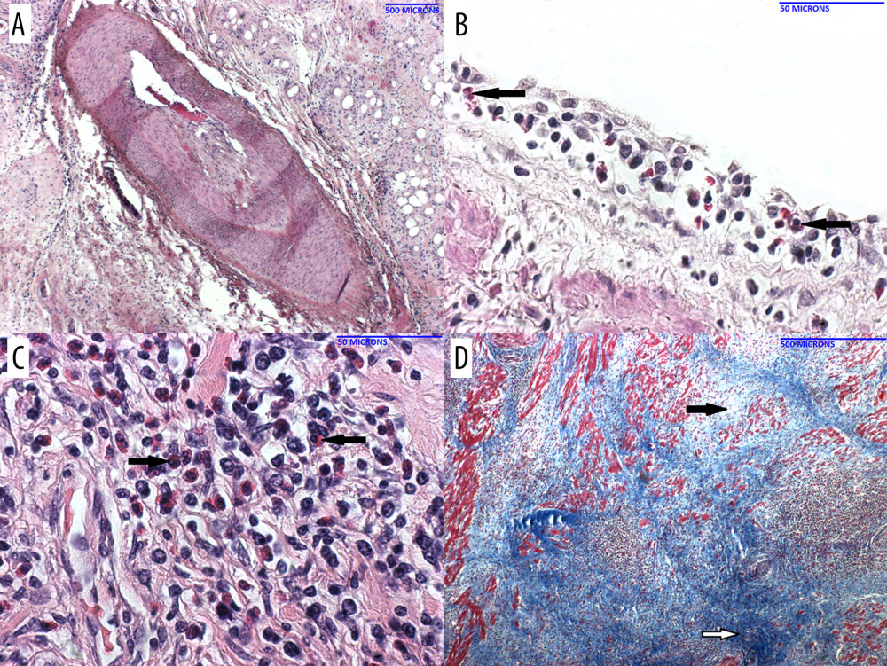
Figure 5 Histopathology of the pig heart (A–D) in B1917 at necropsy(A) Coronary artery with layers of intimal hyperplasia, suggesting prior episodes of luminal thrombosis followed by organization. (B) Veins showed subendothelial (and perivascular) inflammation that contained a mixed inflammatory infiltrate that included eosinophils (arrow). (C) The myocardial interstitium showed areas of mixed inflammation including eosinophils (arrows). (D) The myocardial interstitium showed evidence of remote scarring (dark blue; white arrow) and more recent scarring (light blue; black arrow) [trichrome stain].