29 September 2020: Original Paper
Surgical Drains After Laparoscopic Donor Nephrectomy: Needed or Not?
Haydar Celasin1BC, Akın Fırat Kocaay2A, Sanem Guler Cimen3DEF*, Suleyman Utku Çelik4ABCF, Nurian Ohri2D, Şule Şengül5EF, Kenan Keven5ABC, Acar Tüzüner1ABCDDOI: 10.12659/AOT.926422
Ann Transplant 2020; 25:e926422


Abstract
BACKGROUND: Routine placement of prophylactic drains after laparoscopic donor nephrectomy has been suggested and has become common practice in some centers. However, there is a lack of evidence proving the surgical benefits of routine drain placement in laparoscopic donor nephrectomy. Here, we assessed the effect of surgical drain placement on recovery, length of hospital stay, and complication rates of live kidney donors.
MATERIAL AND METHODS: This retrospective study included all live donor nephrectomies performed at a single institution from January 2010 to January 2017. Surgeries were performed by 2 surgeons; one routinely placed a closed suction drain after LDN whereas the other did not. Patients operated on by these 2 surgeons were enrolled in either the drain or no drain group. Demographic data, preoperative and postoperative creatinine levels, estimated blood loss (EBL), surgical time, surgical complications, and length of hospital stay were compared.
RESULTS: The study included 272 patients. Three were converted to open donor nephrectomy and were excluded (1.1%). Among the 269 patients, 156 (57.9%) had surgical drains and 113 (42.1%) did not. Mean surgical time, estimated blood loss, and duration of hospital stay did not significantly differ between groups. Postoperative complications were encountered in 17 of the patients, but the overall complication rate did not differ between patients with vs. those without surgical drains.
CONCLUSIONS: There was no significant difference between the drain and no drain groups in terms of length of hospital stay, complication rates, or postoperative creatinine levels. Thus, placement of a surgical drain in the setting of an LDN is not justified based on our single-center experience.
Keywords: Directed Tissue Donation, Drainage, Kidney Transplantation, Laparoscopy, Living Donors, Nephrectomy, operative time, Postoperative Complications, Tissue and Organ Harvesting
Background
The current organ shortage is a major challenge in organ transplantation [1]. To overcome this challenge, living organ donation is increasingly accepted and living donor kidney transplantation has become the first and best choice of treatment for patients with end-stage renal disease (ESRD) [1]. Additionally, advancements in immunology, improvements in surgical techniques, and increasing experience with transplantations have significantly decreased the graft loss rate. With these accomplishments, 1-year patient and graft survival have reached 95% [2].
However, clinical studies on organ donors remain scarce. The donor surgery is a unique intervention performed on a healthy volunteer; therefore, its outcomes should be studied separately. When possible, minimally invasive procedures should be preferred to reduce postoperative pain and hospital stay and to improve quality of life of living donors [3]. A laparoscopic donor nephrectomy (LDN) with small extraction site is the criterion standard that provides the most comfort to living donors.
However, safety and comfort does not go hand in hand at all times. Particularly in LDN, every effort should be made to assure a balance between safety and comfort. While living kidney donation appears to be relatively low risk and comfortable for the donor, there are anecdotal and published reports of serious complications and even death attributed to LDN [4]. These complications were mainly due to postoperative hemorrhage, chyle leak, or iatrogenic injury to the bowel.
To detect these complications earlier, routine placement of prophylactic drains after LDN has been suggested and has become common practice in some centers. This practice was based on experience with other abdominal surgeries, such as laparoscopic cholecystectomy, splenectomy or colectomy [5,6]. In these surgeries, the drains have helped surgeons to identify postoperative hemorrhage, as well as to drain or monitor residual intra-peritoneal fluids like bile, fecal material, and pancreatic juice. However, there is a lack of evidence proving the surgical benefits of routine drain placement in LDN. Additionally, routine use of surgical drains may compromise post-surgical comfort.
In this retrospective clinical study, we investigated the influence of surgical drain placement on the recovery, hospital stay, and complication rates of live donors who underwent LDN.
Material and Methods
SURGICAL TECHNIQUE:
The position of the donor was lateral decubitus in all cases. All patients received 1 g of Cefazolin for prophylaxis. The first trocar was inserted peri-umbilically, and the abdomen was insufflated with CO2. A video-endoscope was introduced, and 3 to 4 additional trocars were inserted, as described in the literature [7]. The right or left hemi-colon was dissected from the lateral abdominal wall and mobilized medially. Gerota’s fascia was opened, and the kidney was dissected from the surrounding connective tissue. The renal vessels and ureter were dissected. Subsequently, an 8-cm Pfannenstiel incision was made while maintaining pneumoperitoneum. The ureter was clipped distally and divided, followed by stapling and division of the renal artery and vein.
Subsequently, the kidney was extracted by hand and cooled immediately. Pneumoperitoneum was re-established, and hemostasis was checked in the operative field. A closed-loop suction drain was placed at the surgical site through one of the 5-mm trocars in the drain group. No drains were placed in the other group. The Pfannenstiel incision was closed by continuously suturing the fascia with no. 1 loop polydioxanone stitch, and skin incisions were closed using skin staplers.
POSTOPERATIVE FOLLOW-UP:
Foley catheters, which were routinely placed in all donors intraoperatively, were removed on the day after surgery. All the drains placed during surgery were closed-suction drains. They were removed when output was less than 30 cc per day with serous discharge.
During the postoperative follow-up, intra-abdominal infections were diagnosed via a computed tomography scan, and hemorrhage was diagnosed via changes in patient hemodynamics of and hemoglobin drop in complete blood counts. We directly monitored hemorrhagic drainage through the drain in the drain group, and retroperitoneal ecchymosis and abdominal distention with discomfort assisted the diagnosis in the no drain group. Urinary tract infections were suspected based on symptoms and were diagnosed by urine analysis and culture.
Symptomatic urinary tract infections were treated with oral antibiotics. Surgical wound infections were treated with daily wound dressings and oral antibiotics when necessary. Hemorrhage was treated, if clinically symptomatic, with packed red blood cell (PRBC) replacement and close monitoring, and intra-abdominal infections or abscesses were drained via ultrasonic guidance and intravenous antibiotics.
STATISTICAL ANALYSIS:
Descriptive statistics were used to present the data. As a measure of central tendency, mean values were calculated. Standard deviation was displayed as the measure of variability. Statistical analysis was performed with SPSS 20.p software using the chi-square test and
Results
The study included 272 adult patients who underwent live donor nephrectomy. The medical records revealed that 269 of these patients underwent a standard LND and 3 (1.1%) were converted to open donor nephrectomy due to intraoperative bleeding. These 3 patients who were converted to open were excluded from the study. The remaining 269 patients underwent standard laparoscopic donor nephrectomy. The mean donor age was 47.6±11.1 years and 112 of the donors were female (43.1%). There was no difference between the 2 groups in terms of preoperative creatinine levels, which were all within normal range.
Among the 269 patients, 156 (57.9%) had surgical drains and 113 (42.1%) did not. Left LDN was performed in 242 (89.9%) donors and 28 donors underwent right LDN (10.1%). The mean surgical time was 68.4±24.5 min and did not significantly differ between the 2 groups. Similarly, intraoperative EBL was 150±30 mL and was not significantly different between groups.
Postoperative creatinine levels and mean duration of hospital stay were not significantly different between the groups. In the drain group, the mean drain duration was 3.35±0.81 days. Age, sex, side of nephrectomy, creatinine values, surgical time, EBL, duration of hospital stay, and postoperative complications are demonstrated in Table 1. Postoperative complications were encountered in 17 (6.3%) patients. The most common complication was intra-abdominal hemorrhage, which was detected in 6 donors. One of these cases had tachycardia and required PRBC transfusion classified as a Clavien-Dindo class II complication (Table 2). This patient belonged to the drain placement group and presented with hemorrhagic discharge from the drain, abdominal distention, tachycardia, and mild hypotension. Clinically symptomatic urinary tract infections requiring antibiotic treatments were seen in 3 patients, 2 in the no drain group and 1 in the drain group. None of the urinary tract infections recurred after treatment. Wound infections were encountered in 4 patients; 3 of them were in the no drain group. There was no significant difference between the 2 groups in terms of wound infections. A total of 4 patients developed intra-abdominal abscesses diagnosed by ultrasound or computed tomography scans. One was located in the pelvis behind the uterus, and the others were at the surgical dissection site. The patient with the pelvic abscess was in the no drain group. All abscesses were drained under local anesthesia by interventional radiology, followed by antibiotic treatment according to the bacterial culture results. No recurrent abscesses were encountered. The overall complication rate did not differ by presence or absence of surgical drains. No patients were taken back to the operating room for any of the listed complications.
Discussion
Kidney transplantation is the criterion standard of treatment for patients with ESRD. Living donation has clear advantages over deceased donor kidney transplantation, and with the continuing organ shortage, it also can reduce the number of patients able to receive a cadaveric organ for transplantation. According to the data provided by the Turkish Ministry of Health, over the last year, 2347 live donor kidney transplantations (78%) were performed [8]. Similarly, the increase of living donation is significant worldwide: last year, 31 924 (46%) kidney transplants were performed from live donors [1]. The major problem with living kidney donation is that a healthy person has to undergo a major surgical procedure to provide the organ for transplantation; therefore, a nephrectomy technique that is associated with the lowest risk for the donor with minimal complication rate and invasiveness should be preferred.
The overall complication rate for standard LDN has been reported to be between 3% and 22% [4,9,10]. These complications include pulmonary emboli, surgical wound infections, intra-abdominal hemorrhage, chyle leak, abscesses, and urinary tract infections. Some of these complications are directly related to the technique and extent of surgical dissection at the renal hilum.
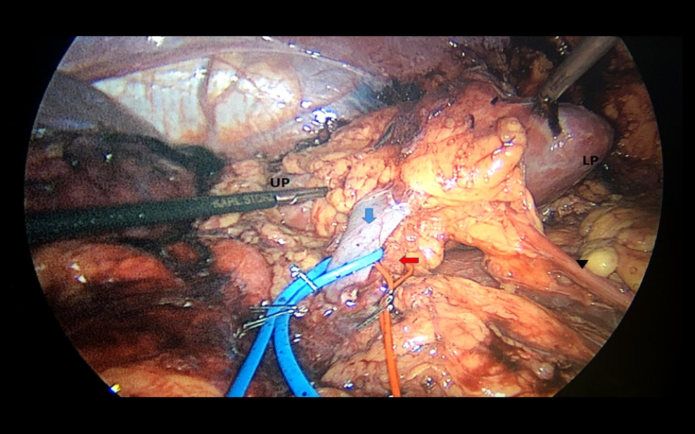
Renal hilar dissection is clearly the most crucial part LDN. Especially on the left side, mapping out the venous anatomy intraoperatively is important due to the relatively high incidence of variations and aberrant lumbar veins. Dissection in this area should be meticulously performed using delicate instruments. Lymphatics are dense around the renal vessels, and division of the perirenal and periaortic lymphatics should be performed with energy devices to reduce the risk of chylous leakage. The renal artery should also be carefully dissected and exposed as proximally as possible. Early branching of the arteries may require more extensive dissection; however, over-dissection and transection should be avoided. The extension of hilar dissection preferred in our center is shown in Figure 1. Using this dissection, we have not encountered any lymphatic leakage-related complications in our series.
In the present study, the postoperative complication rate was 6.4% and did not differ according to placement of surgical drains. However, 6 cases had postoperative hemorrhage and 1 of these patients required PRBC transfusions.
Although these hemorrhages were diagnosed with the help of an existing drain in the drain group, it was not overlooked in the no drain group and was diagnosed promptly with changes in vital signs and hemoglobin levels. Accordingly, no significant difference was found between the 2 groups in terms of complication rates.
Prophylactic placement of drains postoperatively has been widely practiced since the mid-1800s, with an attitude of ’When in doubt, drain’. Since then, several clinical studies evaluated the pros and cons of the use of surgical drains [11–14]. Some studies found a direct correlation between the use of surgical drains and intra-abdominal infections, including surgical wound infections, abdominal pain, and diminished pulmonary functions leading to a prolonged hospital stay [13,14–16]. Furthermore, Lubawski et al. found that the use of drains was associated with postoperative ileus and delayed return of bowel functions after surgery [17]. Additionally, Laine et al. suggested that the use of drains might trigger infections by disseminating bacterial flora in a retrograde fashion, cause ascites formation due to peritoneal irritation and inducing abdominal pain [18]. Consequently, they emphasized the futility of drain usage and recommended drain use only in pancreatic surgeries and emergent cases.
At the present time, little information exists in the literature about the use of drains after laparoscopic surgeries. In the era of laparoscopic surgery, more studies should be performed to eliminate dogmatic surgical practices originating from open surgeries of the past. In our study, drains were not found to be associated with safer outcomes. However, routine placement of surgical drains after LDN might increase the cost of this surgery. Unfortunately, no cost analysis was performed to compare the 2 groups in this study. Future studies should focus on randomized placement of surgical drains after LDN and compare the groups in terms of cost-benefit ratio as well as surgical outcomes and satisfaction rates of the donors.
Conclusions
To the best of our knowledge, this study is the first to analyze the role of surgical drains and their impact on outcomes after LDN. Our findings revealed there was no significant difference between the drain and no drain groups in terms of hospital stay, complication rates, or postoperative creatinine levels. Because donor nephrectomy is a surgery for a patient who does not have any disease and is volunteering to donate a kidney, drain placement can be justified when an intraoperative necessity arises to ensure the safety of the donor. In our experience, in case of postoperative bleeding, the diagnosis can be made based on clinical presentation, changes in vital signs, and radiological imaging, if needed. Thus, non-placement of a surgical drain in the setting of an LDN case is justified based on our single-center experience.
References
1. Organ Procurement and Transplantation Network (OPTN), Scientific Registry of Transplant Recipients (SRTR): OPTN/SRTR 2012 Annual Data Report, 2014, Rockville, MD, Department of Health and Human Services, Health Resources and Services Administration, Healthcare Systems Bureau, Division of Transplantation
2. Cimen S, Guler S, Tennankore K, Surgical drains do not decrease complication rates but are associated with a reduced need for imaging after kidney transplant surgery: Ann Transplant, 2016; 21; 216-21
3. Shockcor NM, Sultan S, Alvarez-Casas J, Minimally invasive donor nephrectomy: Current state of the art: Langenbecks Arch Surg, 2018; 403(6); 681-91
4. Lentine KL, Lam NN, Axelrod D, Perioperative complications after living kidney donation: A national study: Am J Transplant, 2016; 16(6); 1848-57
5. Petrowsky H, Demartines N, Rousson V, Clavien PA, Evidence-based value of prophylactic drainage in gastrointestinal surgery: A systematic review and meta-analyses: Ann Surg, 2004; 240(6); 1074-85
6. Zhang W, He S, Cheng Y, Prophylactic abdominal drainage for pancreatic surgery: Cochrane Database Syst Rev, 2018; 6; CD010583
7. Ratner LE, Ciseck LJ, Moore RG, Laparoscopic live donor nephrectomy: Transplantation, 1995; 60(9); 1047-49
8. : Republic of Turkey, Ministry of Health Transplantation Statistics Retrieved from https://organ.saglik.gov.tr/ContentView.aspx?q=1
9. Halgrimson WR, Campsen J, Mandell MS, Donor complications following laparoscopic compared to hand-assisted living donor nephrectomy: An analysis of the literature: J Transplant, 2010; 2010 825689
10. Lentine KL, Patel A, Risks and outcomes of living donation: Adv Chronic Kidney Dis, 2012; 19(4); 220-28
11. Fong Y, Brennan MF, Brown K, Drainage is unnecessary after elective liver resection: Am J Surg, 1996; 171(1); 158-62
12. Gurusamy KS, Samraj K, Routine abdominal drainage for uncomplicated open cholecystectomy: Cochrane Database Syst Rev, 2007(2); CD006003
13. Merad F, Yahchouchi E, Hay JM, Prophylactic abdominal drainage after elective colonic resection and suprapromontory anastomosis: A multicenter study controlled by randomization. French Associations for Surgical Research: Arch Surg, 1998; 133; 309-14
14. Alvarez Uslar R, Molina H, Total gastrectomy with or without abdominal drains. A prospective randomized trial: Rev Esp Enferm Dig, 2005; 97; 562-69
15. Monson JR, Guillou PJ, Keane FB, Cholecystectomy is safer without drainage: The results of a prospective, randomized clinical trial: Surgery, 1991; 109; 740-46
16. Benedetti-Panici P, Maneschi F, Cutillo G, A randomized study comparing retroperitoneal drainage with no drainage after lymphadenectomy in gynecologic malignancies: Gynecol Oncol, 1997; 65; 478-82
17. Lubawski J, Saclarides T, Postoperative ileus: Strategies for reduction: Ther Clin Risk Manag, 2008; 4(5); 913-17
18. Laine M, Mentula P, Koskenvuo L, When should a drain be left in the abdominal cavity upon surgery?: Duodecim, 2017; 133(11); 1063-68
Tables
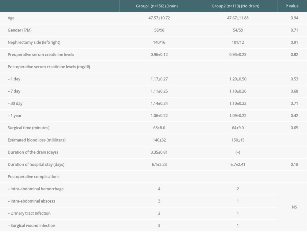 Table 1. Demographic data, serum creatinine levels, surgical time, duration of hospital stay, and postoperative complication rates of the drain and no drain groups.
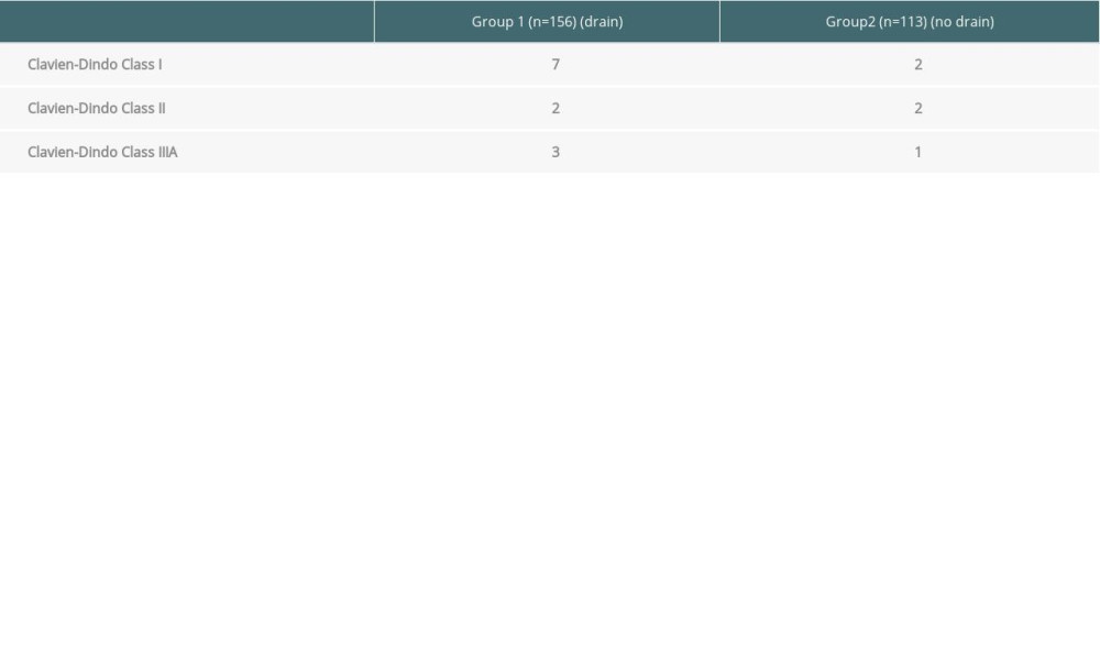 Table 2. Classification of postoperative complications after laparoscopic donor nephrectomy according to Clavien-Dindo system.Table 1. Demographic data, serum creatinine levels, surgical time, duration of hospital stay, and postoperative complication rates of the drain and no drain groups.Table 2. Classification of postoperative complications after laparoscopic donor nephrectomy according to Clavien-Dindo system.
 In Press
Case report
Tongue Carcinoma in Immunosuppressed Patients After Liver and Kidney Transplantation: A Case SeriesAnn Transplant In Press; DOI: 10.12659/AOT.951715
Original article
Prevalence and Risk Factors of Hepatic Steatosis in Kidney Transplant RecipientsAnn Transplant In Press; DOI: 10.12659/AOT.952251
Original article
The Anatomical Landscape of Living Donor Livers: A 101-Case Retrospective Single-Center Study in Indonesia ...Ann Transplant In Press; DOI: 10.12659/AOT.952031
Original article
Decreased Ventilation Duration and ICU Stay Associated With Early Percutaneous Dilatational Tracheostomy Af...Ann Transplant In Press; DOI: 10.12659/AOT.953143
Most Viewed Current Articles
24 Aug 2021 : Review article 20,545
Normothermic Machine Perfusion (NMP) of the Liver – Current Status and Future PerspectivesDOI :10.12659/AOT.931664
Ann Transplant 2021; 26:e931664
29 Dec 2021 : Original article 16,641
Efficacy and Safety of Tacrolimus-Based Maintenance Regimens in De Novo Kidney Transplant Recipients: A Sys...DOI :10.12659/AOT.933588
Ann Transplant 2021; 26:e933588
05 Apr 2022 : Original article 15,898
Impact of Statins on Hepatocellular Carcinoma Recurrence After Living-Donor Liver TransplantationDOI :10.12659/AOT.935604
Ann Transplant 2022; 27:e935604
22 Nov 2022 : Original article 15,796
Long-Term Effects of Everolimus-Facilitated Tacrolimus Reduction in Living-Donor Liver Transplant Recipient...DOI :10.12659/AOT.937988
Ann Transplant 2022; 27:e937988