


01 September 2026: Original Paper
Protective Effects of Quercetin and Sucrose on Cold Preservation Injury in Porcine Kidneys Donated After Cardiac Death
Daisuke Ishii BCDEF 1,2, Asuka Toriumi BCDEF 1, Hiromichi Obara  ABCE 3, Yoshiyasu Satake BC 1, Yoko Okada BC 2, Haruka Dewa G 4, Masashi Imamura G 4, Naoto Matsuno ABCDEG 1,2*
DOI: 10.12659/AOT.952503
Ann Transplant 2026; 31:e952503


Abstract
BACKGROUND: The University of Wisconsin (UW) solution is the gold standard for kidney preservation. Quercetin (QE), a widely used flavonoid, possesses strong antioxidant, anti-inflammatory, and anti-apoptotic properties. Sucrose (Suc) has shown beneficial effects as a supplement. The aim of this study was to investigate the protective effects of QE and Suc against cold injury in porcine kidneys and to evaluate whether their use improves ischemia-reperfusion (I/R) injury after simple CS.
MATERIAL AND METHODS: Kidney grafts were procured after 30 min of warm ischemia, followed by preservation under 2 conditions for 22 h: Group 1, preserved with CS/UW solution (n=4), and Group 2, preserved with CS/UW solution containing QE 33.1 μM and Suc 0.1 M (n=6). All grafts were evaluated using an ex vivo isolated kidney reperfusion model with oxygenated, diluted autologous blood at room temperature.
RESULTS: At the end of reperfusion, creatinine phosphokinase and lactate dehydrogenase levels were significantly lower in Group 2 than in Group 1. We observed more vacuolation in the tubular epithelial cell in Group 1 than in Group 2, and the tubular epithelium showed more irregularity and exfoliation in Group 1 than in Group 2. Additionally, Group 2 had a significantly higher number of anti-ERG-positive endothelial cell nuclei than Group 1 at the end of reperfusion.
CONCLUSIONS: Our results suggest that the addition of QE and Suc to UW solutions can effectively prevent cold injury in DCD kidneys, with QE exerting this beneficial action, possibly by suppressing oxidative stress, and could potentially enhance kidney transplantation outcomes.
Keywords: kidney, Nephrology, Organ Preservation, Transplantation
Introduction
Shortages of organ donors is a universal problem in transplantation. The University of Wisconsin (UW) solution has been the gold standard for preserving the liver, kidneys, and pancreas since it was first developed by Belzer and Southard [1]. Quercetin (QE), a widely used flavonoid, possesses strong antioxidant [2], anti-inflammatory [3], and anti-apoptotic properties [4]. Some bioflavonoids have been reported to attenuate renal tubular cell injury during cold preservation in EuroCollins and UW solutions [5]. In addition, the beneficial effects of the preservation solution may be due to its ability to prevent tissue edema during cold storage (CS). Sucrose (Suc) has shown beneficial effects as a supplement [6]. The aim of this study was to investigate the protective effects of QE and Suc against cold injury in porcine kidneys and to evaluate whether their use improves ischemia-reperfusion (I/R) injury after simple CS. This study is investigating the mechanisms underlying the efficacy of QE and Suc by utilizing cytokines and immune tissues. We believe it will contribute to the development of new, more effective preservation solutions.
Material and Methods
ANIMALS:
The Institutional Animal Ethics Committee of the Clinical Research Center, Asahikawa Medical University, Japan (permit no. R4-089) approved all experimental procedures. We used domestic pigs (male, crossbred Large-Yorkshire, Landrace, and Duroc hogs, 2–3 months old, 30 kg; Taisetsu Sanroku-Sya, Asahikawa, Japan) in this study.
Midazolam (0.3 mg/kg), medetomidine hydrochloride (0.06 mg/kg), and butorphanol tartrate (0.03 mg/kg) were injected into porcine femoral muscle for anesthetic induction. The peripheral ear vein was cannulated, and Ringer’s lactate solution was infused at a rate of 6.7 ~ 10 ml/kg/hr. Thiamylal sodium (4.2 mg/kg) was intravenously injected and intubation was performed. Anesthesia was maintained with propofol and oxygen (4 L/min). The internal jugular vein was then infused with 6% (w/v) hydroxyethyl starch (33.3 ml/kg/hr).
The domestic pigs underwent midline laparotomy. The right common iliac artery was isolated and a catheter was inserted to procure ~ 400 mL of autologous whole blood. The kidney ureter, artery, and vein were identified, and the blood flow cut off. We intravenously infused potassium chloride (2 mEq/kg) and cardiac arrest was induced. The kidneys were kept under warm ischemic time (WIT) conditions for 30 min which simulated the kidney donation after circulatory death (DCD). The kidney graft was retrieved and washed from the renal artery with 200 mL of each preservation solution at 4 °C as a back-table procedure. The kidney grafts were preserved under CS for 22 hours at 4 °C.
STUDY DESIGN:
The kidney grafts procured after 30 min of WIT were preserved under 2 conditions for 22 hours: Group 1, preserved with UW solution (n = 4) and Group 2, preserved with UW solution containing QE 33.1 μM and Suc 0.1 M (n = 6). Quercetin was dissolved in dimethyl sulfoxide (DMSO) to a concentration of 100 mg/mL. One milliliter of this solution, diluted to 10 mg/mL with DMSO, was dissolved in 1 L of deionized water to achieve a concentration of 33.1 μM.
ISOLATED EX VIVO REPERFUSION MODEL:
Kidney function was evaluated using an isolated kidney reperfusion model. The grafts were then reperfused with oxygenated diluted autologous blood at room temperature. Reperfusion of the renal artery was set at 90–100 mm Hg and ~ 20 ml/min. The reperfusion fluid was autologous whole blood containing 500 mL saline, 250 mL dextran, 60 mL calcium gluconate hydrate, 22 mL 8.5% bicarbonate, and heparin. Heparin levels were adjusted to an activated clotting time of 300 s. The hematocrit was maintained at ~ 10–12%. The oxygenator was regulated to achieve physiological blood gas values (pO2, ~ 290–300 mm Hg; pCO2, 30–50 mm Hg).
BIOCHEMISTRY:
Blood urea nitrogen (BUN), creatinine (Cr), creatinine phosphokinase (CPK), and lactate dehydrogenase (LDH) levels in the perfusate were measured every 60 min during reperfusion to determine the viability of the preserved kidney grafts using standard biochemical methods. In addition, lactate levels were measured every 60 min during reperfusion using a blood gas analyzer (i-STAT 1 analyzer; Abbott Japan LLC, Tokyo, Japan).
HISTOPATHOLOGICAL EVALUATION:
A kidney wedge biopsy was performed immediately after laparotomy, before and after preservation, and 1 and 3 h after isolated ex vivo reperfusion. The kidney tissue was fixed in 10% phosphate-buffered formalin and embedded in paraffin. Each sample was sectioned into 3 μm slices, dehydrated using ethanol, and hematoxylin-eosin staining was performed.
ERG immunohistochemistry was performed to assess endothelial cell damage in glomerular and peritubular capillary networks. Additionally, CD42b immunohistochemistry was performed to assess platelet aggregation in the glomerular tufts and around the renal tubules.
A primary rabbit anti-ERG monoclonal antibody was used, obtained from Nichirei Bioscience Co.,
Ltd. (clone EP111; Tokyo, Japan) and a rabbit anti-CD42b polyclonal antibody from the Proteintech Group, Inc. (GeneID 2811; IL, United States). Envision-labeled polymer reagent (DAKO, Glostrup, Denmark) was used as the secondary antibody. ImageJ (U.S. National Institute of Health, Bethesda) was used to quantify the anti-ERG and CD42b positive area. ImageJ software was used, obtainable from
GENE EXPRESSION:
Quantitative reverse transcription polymerase chain reaction was used to determine the gene expression of endothelial-specific, inflammatory, and apoptotic proteins. Renal parenchyma biopsies were performed after laparotomy, before and after preservation, and 3 hours after reperfusion. The samples were stored at −80 °C until analysis. Total RNA was extracted from snap-frozen kidney biopsies using an miRNeasy Micro Kit (Qiagen, Valencia, CA, USA). RNA concentration was determined using a spectrophotometer (NanoDrop 2000; NanoDrop Technologies, Wilmington, DE, USA). Equal amounts of RNA were converted to complementary DNA using the Transcriptior First Standard cDNA Synthesis Kit (Roche, Basel, Switzerland), and complementary DNA levels were measured using a LightCycler 480 System II (Roche, Basel, Switzerland). The relative expression of the mRNA of interest was normalized to the housekeeping gene—glyceraldehyde-3-phosphate dehydrogenase (GAPDH). Data are presented as the relative quantification of GAPDH. Table 1 summarizes sense and antisense primer sequences.
STATISTICAL ANALYSES:
We presented the results as the mean ± standard deviation. Statistical analyses were performed using Microsoft Excel 2013 (Microsoft Corp., Redmond, WA, USA). The results were analyzed using the
Results
BUN, CR, CPK, LDH, AND LACTATE IN THE PERFUSATE SOLUTION:
No BUN level differences were observed between the groups. At the end of reperfusion, the BUN levels in Groups 1 and 2 were 4.30 ± 0.56 and 4.65 ± 1.01 mg/dl, respectively (P = 0.598) (Figure 1A). Cr levels did not differ between groups. At the end of reperfusion, the Cr levels in Groups 1 and 2 were 0.45 ± 0.07 and 0.49 ± 0.45 mg/dl, respectively (P = 0.431) (Figure 1B).
At 0, 1, and 2 h, and at the end of reperfusion, CPK levels were significantly lower in Group 2 at 313.16 ± 171.12, 391.00 ± 184.33, 449.33 ± 189.95, and 504.66 ± 200.33 IU/L, respectively, than in Group 1 at 1249.75 ± 188.50, 1409.25 ± 204.43, 1490.25 ± 149.65, and 1504.00 ± 94.92 IU/L, respectively (Group 2 vs Group 1; P < 0.005, P < 0.005, P < 0.005, P < 0.005) (Figure 1C).
At 0, 1 h, and the end of reperfusion, the LDH levels were significantly lower in Group 2 than in Group 1, at 154.16 ± 25.54 vs 215.55 ± 35.42, 233.33 ± 61.90 vs 411.00 ± 101.88, and 573.66 ± 151.52 vs 836.00 ± 141.08 mg/dl (Group 2 vs Group 1; P < 0.05, P < 0.05, P < 0.05). The LDH levels at 2 h were not different between the groups (634.00 ± 166.12 [Group 1] vs 420.50 ± 141.77 mg/dl [Group 2]; P = 0.080) (Figure 1D). No differences were observed in lactate levels between the groups. At the end of reperfusion, the lactate levels in Groups 1 and 2 were 10.84 ± 0.86 and 10.38 ± 0.69 mg/dl, respectively (P = 0.611) (Figure 1E).
HISTOLOGICAL FINDINGS DURING REPERFUSION:
Hematoxylin-eosin staining revealed that at the end of reperfusion, there was more vacuolation in tubular epithelial cells in Group 1 than in Group 2. In addition, the tubular epithelium showed more irregularity and exfoliation in Group 1 than in Group 2. In Group 1, there was more interstitial inflammation and hemorrhaging than in Group 2 (Figure 2A, 2B).
ERG AND CD42B IMMUNOHISTOCHEMISTRY:
At the end of reperfusion, Group 2 had significantly higher numbers of anti-ERG staining-positive endothelial cell nuclei than Group 1 (433.70 ± 151.69 [Group 2] vs 378.02 ± 119.29 [Group 1] per field; P = 0.046) (Figure 3A–3C). However, the positive areas of anti-CD42b staining at the end of reperfusion were not different between the groups (22087.62 ± 14258.80 [Group 1] vs 17176.51 ± 10944.07 [Group 2] per field; P = 0.073) (Figure 4A–4C).
GENE EXPRESSION:
There were no differences in gene expression between groups. At the end of reperfusion, Groups 1 and 2 did not differ in the expression of the tumor necrosis factor (TNF-α) (0.2223 ± 0.0749 vs 0.1423 ± 0.0579; P = 0.108; Figure 5A), interferon-γ (IFN-γ) (0.0797 ± 0.0406 vs 0.0447 ± 0.0183; P = 0.096; Figure 5B), and caspase-3 (cas-3) (0.4381 ± 0.1785 vs 0.3269 ± 0.1156; P = 0.354; Figure 5C).
Discussion
Conclusions
We investigated the utility of flavonoids QE and Suc in kidney transplantation using a reperfusion model in CS. Our results suggest that the addition of QE and Suc to UW solutions can effectively prevent cold injury in DCD kidneys, with QE exerting this beneficial action, possibly by suppressing oxidative stress, and could potentially enhance kidney transplantation outcomes.
Figures
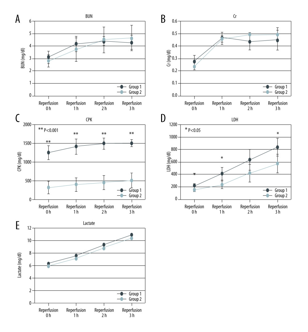 Figure 1. Laboratory test results. (A) BUN (mg/dl), (B) Cr (mg/dl), (C) CPK (IU/L), (D) LDH (mg/dl), and (E) lactate (mg/dl) levels (mean ± SD) in the perfusate solution before, and 1, 2, and 3 h after reperfusion.
 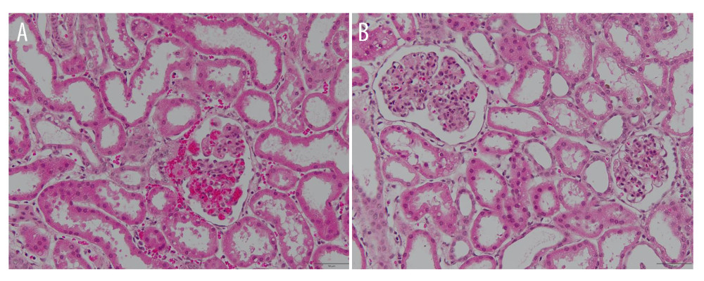 Figure 2. Hematoxylin-eosin staining at the end of reperfusion. (A) Group 1. (B) Group 2.
 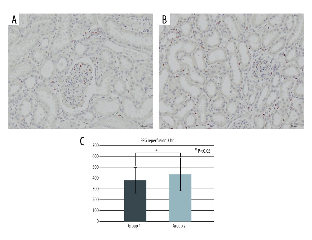 Figure 3. ERG immunohistochemistry at the end of reperfusion. (A) Group 1. (B) Group 2. (C) Quantification of anti-ERG positive area in Groups 1 and 2.
 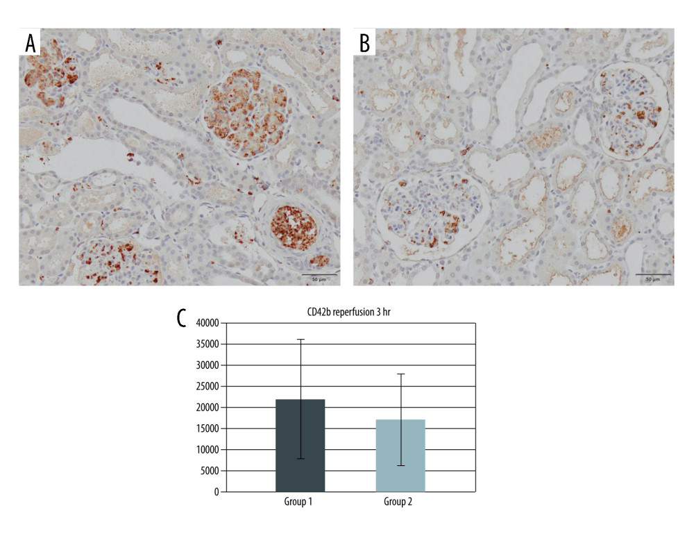 Figure 4. CD42b immunohistochemistry at the end of reperfusion. (A) Group 1. (B) Group 2. (C) Quantification of anti-CD42b positive area in Groups 1 and 2.
 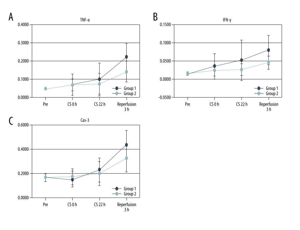 Figure 5. Polymerase chain reaction. (A) TNF-α, (B) IFN-γ, and (C) caspase-3 value after laparotomy, before and after preservation, and 3 h after reperfusion.
 Tables
Table 1. PCR primers.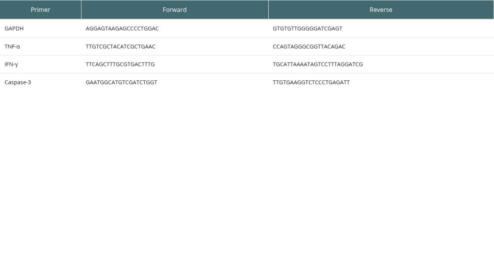
References
1. Southard JH, Belzer FO, Organ preservation: Annu Rev Med, 1995; 46; 235-47
2. Liua C, Zhenga Y, Lua J, Quercetin protects rat liver against lead-induced oxidative stress and apoptosis: Environ Toxicol Pharmacol, 2010; 29(2); 158-66
3. Zielińska M, Kostrzewa A, Ignatowicz E, Budzianowski J, The flavonoids, quercetin and isorhamnetin 3-O-acylglucosides diminish neutrophil oxidative metabolism and lipid peroxidation: Acta Biochim Pol, 2001; 48(1); 183-89
4. Kinaci MK, Erkasap N, Kucuk A, Tosun M, Effects of quercetin on apoptosis, NF-κB and NOS gene expression in renal ischemia/reperfusion injury: Exp Ther Med, 2012; 3(2); 249-54
5. Ahlenstiel T, Burkhardt G, Köhler H, Bioflavonoids attenuate renal proximal tubular cell injury during cold preservation in Euro-Collins and University of Wisconsin solutions: Kidney Int, 2003; 63(2); 554-63
6. Kumano K, Wang W, Endo T, Effects of osmotic agents for cold kidney preservation: Nihon Hinyokika Gakkai Zasshi, 1994; 85(6); 925-31
7. Bertuzzo VR, Cescon M, Odaldi F, Actual risk of using very aged donors for unselected liver transplant candidates; A European single-center experience in the MELD era: Ann Surg, 2017; 265(2); 388-96
8. Flores A, Asrani SK, The donor risk index: A decade of experience: Liver Transpl, 2017; 23(9); 1216-25
9. Hoyer DP, Paul A, Gallinat A, Donor information-based prediction of early allograft dysfunction and outcome in liver transplantation: Liver Int, 2015; 35(1); 156-63
10. Agopian VG, Petrowsky H, Kaldas FM, The evolution of liver transplantation during 3 decades: Analysis of 5347 consecutive liver transplants at a single center: Ann Surg, 2013; 258(3); 409-21
11. Elahi MM, Kong YX, Matata BM, Oxidative stress as a mediator of cardiovascular disease: Oxid Med Cell Longev, 2009; 2(5); 259-69
12. Kaçmaz A, Polat A, User Y, Octreotide: A new approach to the management of acute abdominal hypertension: Peptides, 2003; 24(9); 1381-86
13. Salahudeen AK, Huang H, Patel P, Jenkins JK, Mechanism and prevention of cold storage-induced human renal tubular cell injury: Transplantation, 2000; 70(10); 1424-31
14. Heijnen CG, Haenen GR, Oostveen RM, Protection of flavonoids against lipid peroxidation: The structure activity relationship revisited: Free Radic Res, 2002; 36(5); 575-81
15. Seok YM, Kim J, Park MJ, Wen-pi-tang-Hab-Wu-ling-san attenuates kidney fibrosis induced by ischemia/reperfusion in mice: Phytother Res, 2008; 22(8); 1057-63
16. Spandou E, Tsouchnikas I, Karkavelas G, Erythropoietin attenuates renal injury in experimental acute renal failure ischaemic/reperfusion model: Nephrol Dial Transplant, 2006; 21(2); 330-36
17. Inal M, Altinişik M, Bilgin MD, The effect of quercetin on renal ischemia and reperfusion injury in the rat: Cell Biochem Funct, 2002; 20(4); 291-96
18. Ahmad N, Pratt JR, Potts DJ, Lodge JP, Comparative efficacy of renal preservation solutions to limit functional impairment after warm ischemic injury: Kidney Int, 2006; 69(5); 884-93
19. Kato F, Gochi M, Kawagoe T, The protective effects of quercetin and sucrose on cold preservation injury in vitro and in vivo: Organ Biology, 2020; 27; 207-15
20. Gochi M, Kato F, Toriumi A, A novel preservation solution containing quercetin and sucrose for porcine kidney transplantation: Transplant Direct, 2020; 6(12); e624
21. Soares BLF, Freitas MAL, Montero EF, Alprostadil attenuates inflammatory aspects and leucocytes adhesion on renal ischemia and reperfusion injury in rats: Acta Cir Bras, 2014; 29(Suppl 2); 55-60
22. Ahlenstiel T, Burkhardt G, Köhler H, Kuhlmann MK, Improved cold preservation of kidney tubular cells by adding bioflavonoids to organ preservation solutions: Transplantation, 2006; 81; 231-39
23. Cardinal H, Dieudé M, Hébert M-J, Endothelial dysfunction in kidney transplantation: Front Immunol, 2018; 9; 1130
24. Jansen MPB, Emal D, Teske GJD, Release of extracellular DNA influences renal ischemia reperfusion injury by platelet activation and formation of neutrophil extracellular traps: Kidney Int, 2017; 91; 352-64
25. Kinsey GR, Li L, Okusa MD, Inflammation in acute kidney injury: Nephron Exp Nephrol, 2008; 109(4); e102-7
26. Dong X, Swaminathan S, Bachman LA, Resident dendritic cells are the predominant TNF-secreting cell in early renal ischemia-reperfusion injury: Kidney Int, 2007; 71(7); 619-28
27. Donnahoo KK, Meng X, Ayala A, Early kidney TNF-alpha expression mediates neutrophil infiltration and injury after renal ischemia-reperfusion: Am J Physiol, 1999; 277(3); R922-29
28. Yang B, Lan S, Dieudé M, Caspase-3 is a pivotal regulator of microvascular rarefaction and renal fibrosis after ischemia-reperfusion injury: J Am Soc Nephrol, 2018; 29(7); 1900-16
Figures
Figure 1. Laboratory test results. (A) BUN (mg/dl), (B) Cr (mg/dl), (C) CPK (IU/L), (D) LDH (mg/dl), and (E) lactate (mg/dl) levels (mean ± SD) in the perfusate solution before, and 1, 2, and 3 h after reperfusion.Figure 2. Hematoxylin-eosin staining at the end of reperfusion. (A) Group 1. (B) Group 2.Figure 3. ERG immunohistochemistry at the end of reperfusion. (A) Group 1. (B) Group 2. (C) Quantification of anti-ERG positive area in Groups 1 and 2.Figure 4. CD42b immunohistochemistry at the end of reperfusion. (A) Group 1. (B) Group 2. (C) Quantification of anti-CD42b positive area in Groups 1 and 2.Figure 5. Polymerase chain reaction. (A) TNF-α, (B) IFN-γ, and (C) caspase-3 value after laparotomy, before and after preservation, and 3 h after reperfusion. In Press
Original article
Analysis of Factors Influencing the Psychological State of Patients After Autologous Liver TransplantationAnn Transplant In Press; DOI: 10.12659/AOT.953645
Original article
Fundamental Strategies and Technical Perspectives in Bile Duct Anastomosis for Living Donor Liver Transplan...Ann Transplant In Press; DOI: 10.12659/AOT.953761
Clinical Research
Retrospective Study of Implementation and Outcomes of a Non-Cryopreserved Autologous Hematopoietic Stem Cel...Ann Transplant In Press; DOI: 10.12659/AOT.954230
Most Viewed Current Articles
24 Aug 2021 : Review article 21,883
Normothermic Machine Perfusion (NMP) of the Liver – Current Status and Future PerspectivesDOI :10.12659/AOT.931664
Ann Transplant 2021; 26:e931664
29 Dec 2021 : Original article 18,136
Efficacy and Safety of Tacrolimus-Based Maintenance Regimens in De Novo Kidney Transplant Recipients: A Sys...DOI :10.12659/AOT.933588
Ann Transplant 2021; 26:e933588
22 Nov 2022 : Original article 16,807
Long-Term Effects of Everolimus-Facilitated Tacrolimus Reduction in Living-Donor Liver Transplant Recipient...DOI :10.12659/AOT.937988
Ann Transplant 2022; 27:e937988
05 Apr 2022 : Original article 16,716
Impact of Statins on Hepatocellular Carcinoma Recurrence After Living-Donor Liver TransplantationDOI :10.12659/AOT.935604
Ann Transplant 2022; 27:e935604